MICROTIA TREATMENT OPTIONS


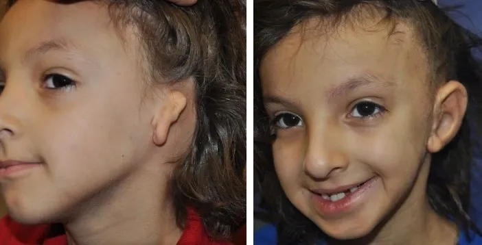
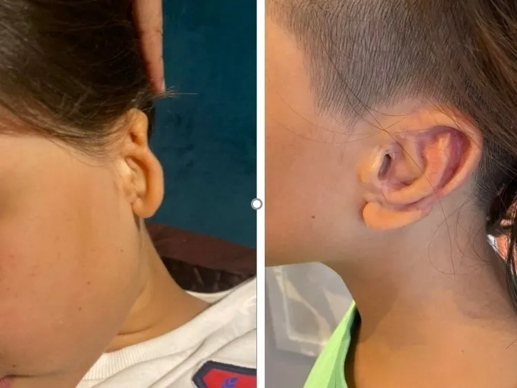
There are currently four treatment options for microtia
- Do nothing
- Rib Graft Reconstruction
- Porous Polyethylene Implant Ear Reconstruction
- Prosthetic Ear
There are also future technologies currently being investigated including 3D printing and tissue engineering.
Each option has advantages & disadvantages. No option is “perfect”, and there are no right or wrong options – just what is best for each individual child and family. It is important for you to be comfortable with whatever option you choose and to collect as much information as possible to help your decision making. You need to wrigh up each option and choose which is right for you and your child.
Ear reconstructive surgery is technically very difficult. Very few surgeons perform ear reconstructions on a regular basis. This is mainly due to the rarity of the condition, but also because of the specific training required. It is therefore important to choose a surgeon who has ongoing experience in this surgery and who can show you pictures of their results.
Do Nothing
There is no medical necessity for ear reconstruction and families can choose not to pursue surgical intervention or wait until their child is older and can make their own decision.
Every child is different and some children have no interest in ear reconstruction and are confident with no psychological or emotional concerns regarding their ear. There are also no time constraints on when to have ear reconstruction. Some patients make the decision to have surgery during adolescence or as an adult, others choose to never have surgery. It is a very personal decision for each child and family.

Rib Graft Ear Reconstruction
The traditional method of ear reconstruction is Rib Graft reconstruction. Rib Graft reconstruction uses tissue from the patients own body and involves making an ear framework from the patients rib cartilage, which is taken from the patient’s rib cage. This technique has been used for the past 50 years. In Australia, this surgery is usually performed in two stages i.e. two separate operations.
During the first surgery, cartilage is harvested from the rib cage. The cartilage from about three ribs is removed and carved into a framework which is designed to resemble the other ear. This procedure leaves a chest scar. A skin pocket is formed, and the cartilage framework is inserted. If there is a lobe present, it is used and repositioned into its normal position.
The second stage of the ear reconstruction, which involves elevation of the created ear from the side of the head, is performed several months after the first stage. Sometimes a child may require surgery to pin back the other ear to more closely match the reconstructed one.
Rib Graft ear reconstructions are usually performed on children 8 -12yrs of age. By this time, the child is usually large enough that rib size is sufficient to harvest the amount of rib cartilage required. If the child is still small, surgery may be postponed until adequate rib for the framework can be harvested.
Polythylene Implant Ear Reconstruction
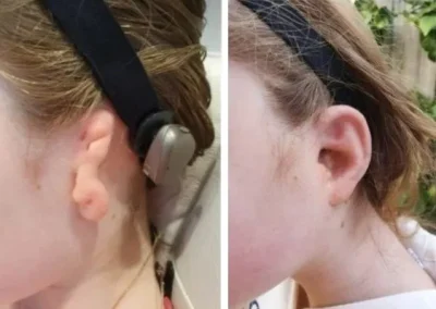
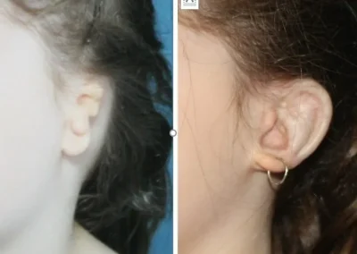
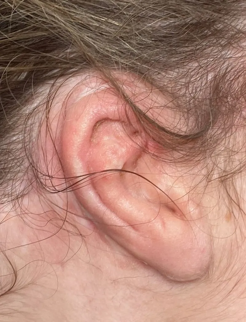
Polyethylene Ear Reconstruction uses a porous synthetic implant and a completely different surgical technique. The implant is placed in position and is then covered with a living membrane which is dissected from the side of the head. The porous material allows blood vessels from the membrane to grow through the implant over time, therefore making it well tolerated by the body. A skin graft is then placed over the membrane, covering the front and back of ear.
This procedure is usually performed in only one surgery and can be done at a younger age – usually from three to four years of age since this method is not dependent on the use of sufficient rib cartilage. This procedure therefore can be performed before the child attends school.
There is often concern about the possibility of rejection of the implant because it is synthetic. Polyethylene is an inert substance and is well tolerated by the body. It has been used in different parts of the body for a long time, and in ear reconstructions overseas since 1991. It is not rejected by the body. But if the implant becomes exposed and the living tissue covering it is damaged, it will need to be replaced.
Originally, this procedure used a two piece implant. This was then improved to use a single piece implant, and now, this procedure can take advantage of new 3D scanning technology to make a custom implant that matches the non-affected ear. Many Australian families have travelled overseas to have this procedure. This procedure is now available in Australia, and was first performed in Sydney in 2019.
Videos
Harriet’s Wish
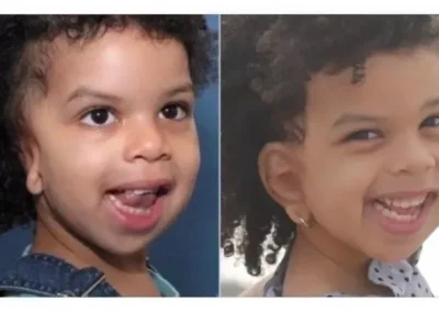
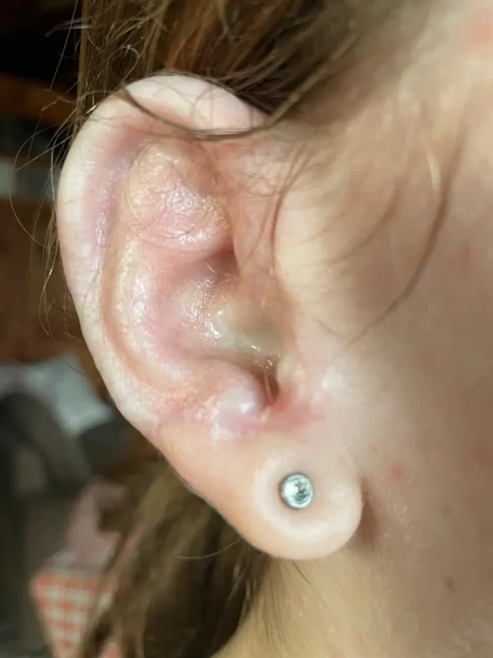
Harriet was born with bilateral microtia and atresia. Her wish is to have two big ears and to wear earrings.
Maddie’s New Ear
Maddie gets a new custom 3D ear implant. She also has an Ossia hearing device implanted during the procedure. Her reaction is priceless when she sees her new ear.
Prosthetic Ear
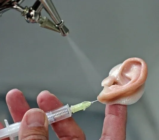
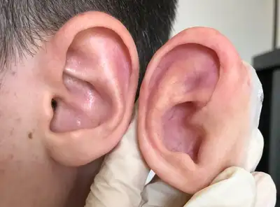
A Prosthetic ear is an artificial ear that is usually made from silicone. The prosthesis can be attached with adhesive or surgically implanted magnets or clips.
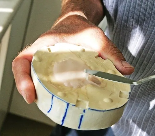
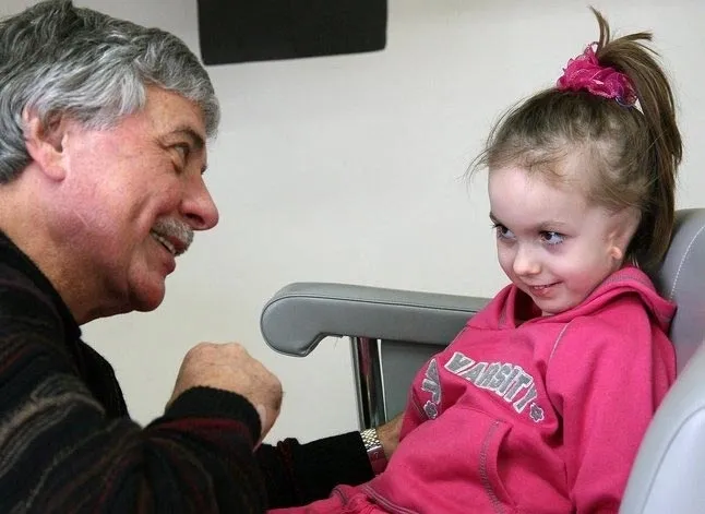
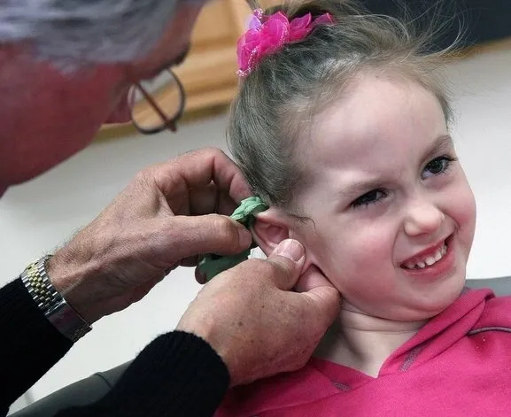
A prosthesis is usually constructed in four steps. The first step is to take an impression of the opposite ear (or a parent’s ear in bilateral cases). Next, a clay sculpture is made of the ear. Then the sculpture is molded and silicone is cast to replicate the ear. Finally, the prosthesis is tinted and hand painted to match the surrounding skin.
This process can create a very realistic-looking ear and has less risk than a surgical reconstruction. It provides an option for patients who are not candidates for surgery.
It can be difficult hiding the seam where the prosthesis meets normal skin and it must be removed at night. Daily cleaning of the prosthesis and skin around the site is required and prosthetic ears need to be replaced every two to three years, so there are ongoing costs.
Future Technologies
There is much interest in the future possibilities for ear reconstruction. Future possibilities include the 3D printing, tissue engineering and the biofabrication of ears. Scanning and 3D anatomical modelling software are now available to generate 3D computer models and customised ear implants for each patient.
3D bioprinted surgically implantable regenerative ears are being researched and investigated. A biodegradable polymer would be printed using 3D biofabricators. Cells and growth factors will be incorporated into the scaffold prior to surgical implantation. It is expected that the polymer scaffold would slowly dissolve, leaving only regenerated tissue.
There are several research projects currently being conducted worldwide, and some clinical trials are also beginning to trial new technologies. It will take some years before these options become readily available for patients, but it is very interesting to follow the progress being made in this area.
Learn more about some of the current research project here:
https://www.youtube.com/watch?v=MD_9q5096zI
https://www.microtia.net/future-of-microtia-surgery/


